
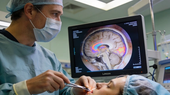
A machine-learning system processed live endoscopic video to help surgeons identify critical anatomy during a pituitary tumor operation, aiming to reduce the risk of vision loss in a high-stakes clinical trial in London
Within the narrow confines of a patient's skull, a neurosurgical team at the National Hospital for Neurology and Neurosurgery in London relied on a machine-learning system to help distinguish vital structures from tumor tissue-while the operation was underway. The stakes were immediate: a misstep could have cost the patient, Rhys Hibbert, his remaining eyesight. Instead, the system processed live endoscopic video, highlighting critical anatomy in real time as surgeons navigated millimeter-scale distances between the tumor, optic nerves, and major blood vessels.
Unlike conventional surgical navigation tools that depend on preoperative scans, the University College London (UCL) software analyzed the actual surgical field as it changed. The system, developed at UCL's Hawkes Institute, was trained on hundreds of annotated videos from previous pituitary operations. This allowed it to recognize not only anatomical features but also surgical instruments and their interactions with tissue, providing a dynamic reference that adapted to the evolving scene inside the patient's brain.
Clinical Trial and Technical Evidence
The clinical trial, funded by the UK's National Institute for Health and Care Research and Google, marked the first time this machine-learning system was used to support live surgery on a human patient. The operation targeted an 11-millimeter pituitary tumor that had already caused severe hormone disruption and progressive vision loss. Hibbert, 48, had lost much of his peripheral vision and required walking sticks before the procedure. Within a week of surgery, he was able to walk unaided and reported significant improvement in sight.
Technical implementation relied on NVIDIA's Clara IGX platform, designed for real-time machine intelligence in medical devices. The system's training dataset comprised hundreds of endoscopic videos, each annotated to identify key anatomical structures and instrument-tissue interactions. This approach exposed the model to a wide range of surgical scenarios, but the trial itself remains limited to a single patient outcome so far. The software acted strictly as a support tool: the lead surgeon, Hani Marcus, retained full control, using the system's visual overlays as an additional reference rather than a replacement for clinical judgment.
Human Oversight and Safety Limits
Pituitary surgery is among the most technically demanding neurosurgical procedures, with critical structures separated by only a few millimeters. Even minor errors can result in blindness, stroke, or death. The UCL system's real-time analysis aimed to reduce the risk of accidental damage by flagging high-risk anatomy as the operation progressed. However, the system did not automate any surgical action or decision. All interventions remained under direct human control, and the technology's role was limited to visual augmentation.
Previous uses of the system were confined to surgical training environments, where annotated video could help residents learn to identify anatomy and avoid common errors. The current trial represents a step toward integrating machine-learning support into live clinical workflows, but the evidence base is still narrow. No independent audit or large-scale evaluation has yet been published, and the system's performance across diverse patient anatomies and surgical teams remains untested. As with other recent advances in real-time robotic and AI-assisted surgery-such as those described in reported earlier-the transition from demonstration to routine practice will require systematic validation and regulatory scrutiny.
Broader Context and Next Steps
Developers at UCL have stated that future versions of the system could track surgical instruments more precisely and monitor their interaction with tissue, potentially offering additional feedback to surgeons during complex procedures. The project has received support from UCLH's Biomedical Research Centre, the Royal College of Surgeons, EPSRC, and Wellcome, reflecting broad institutional interest in machine-learning applications for surgical safety. However, the current evidence is limited to a single successful case, and the system's ability to generalize across different surgical environments, patient anatomies, and operator skill levels remains unproven.
For now, the technology's main contribution is as a real-time visual aid, not as an autonomous surgical agent. The trial's outcome demonstrates that machine-learning systems can be integrated into high-risk clinical workflows without removing human oversight. But the absence of large-scale, independently verified results means that claims of risk reduction or improved outcomes must be treated with caution. The next phase will require rigorous, multi-center trials and transparent reporting of both successes and failures before such systems can be considered for routine deployment in neurosurgery or other high-stakes medical fields.
Machine-learning support in surgery depends on computer vision: algorithms trained to recognize anatomical structures, instruments, and tissue interactions from video data. Unlike static preoperative scans, real-time computer vision must handle variable lighting, occlusion, blood, and unpredictable movement. Training such systems requires large, annotated datasets and careful validation to avoid overfitting to specific cases. Even with high accuracy in controlled settings, real-world deployment introduces new sources of error and uncertainty. Human oversight remains essential, both to interpret the system's output and to intervene when the software fails to recognize rare or ambiguous anatomy. As machine-learning tools move from training labs to operating rooms, the challenge is not only technical performance but also ensuring that safety, accountability, and clinical judgment remain at the center of patient care.