
MIT engineers have built a paper-thin biohybrid robot that swims through water using light-responsive muscle cells. Its maze test demonstrates controlled locomotion with minimal biological material, but not autonomous operation.
A beam of light can steer MIT's paper-thin swimming robot through water because its fins are lined with living muscle tissue. The laboratory prototype followed a manually moved light source through an underwater maze, demonstrating controlled locomotion with far less biological material than earlier three-dimensional biohybrid machines. MIT presented the work on September 29, 2026, describing it as a new aquabot platform published in Advanced Functional Materials. The study is summarized in an MIT research report.
The result is technically precise but easy to oversell. This is not an autonomous underwater robot: researchers moved the light above the robot and controlled its turns by choosing which fin to stimulate. The machine's significance lies in its construction and efficiency rather than in independent navigation. MIT describes it as the first very thin, two-dimensional muscle-powered robot demonstrated to achieve locomotion, with the emphasis on producing useful thrust from a minimal amount of living material.
Light-controlled movement
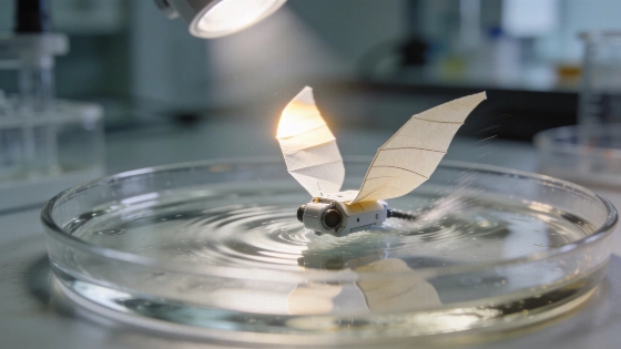
The robot is built around a gelatin-based film approximately 0.5 millimeters thick and roughly comparable in length and width to a piece of chewing gum. Two flexible sections form its fins, and each fin carries a layer of living skeletal muscle cells thinner than a human hair. The cells were genetically engineered to contract when exposed to light, allowing illumination to function as an external control signal without placing conventional motors or a battery on the thin body.
Illuminating one side makes its muscle cells twitch and flap the corresponding fin. That movement pushes against the surrounding water. Switching the light between the two sides changes the robot's direction, while adjusting the timing of illumination changes its speed. The system therefore behaves like two independently addressable biological actuators: lighting one side moves one fin, while lighting both sides activates both fins at once.
In testing, the robot was submerged in a large petri dish containing a simple maze. A researcher manually moved the light source overhead, and the robot followed the illuminated direction through the water. At its fastest measured speed, it traveled approximately four times its own body length in one minute. That body-length-normalized value is the reported ceiling for the present laboratory version. It is considerably slower than a human swimmer, but speed is not the central claim: the demonstration shows that a very thin two-dimensional structure can convert muscle contraction into useful propulsion.
Material determines force
The design extends work by Ritu Raman, an associate professor of mechanical engineering at MIT, and her research group. In an earlier experiment, the team grew muscle cells on a gel disk patterned with concentric and radial grooves. Light-induced contractions stretched and squeezed the disk, but the displacement was limited to approximately 100 micrometers.
A swimming machine required more coordinated force. The researchers therefore treated the supporting material as part of the actuator rather than as a passive scaffold. They tested different gel compositions, stiffness levels and groove geometries to improve how the cells aligned and contracted. This is a central principle of soft robotics: the substrate determines how biological deformation is transmitted, distributed and converted into movement.
Square-bottomed grooves aligned the cells more effectively than curved grooves. That alignment helped the cells fuse into muscle fibers capable of more coordinated contractions. The team also replaced fibrin, an ultrasoft gel that deformed under muscle force, with gelatin methacrylate, or GelMA. Stiffer GelMA formulations supported stronger alignment and contractions while a film about 0.5 millimeters thick remained flexible enough to move with the tissue.
The researchers also repeatedly stimulated the muscle with light before assembling the robot. This training routine strengthened the tissue and improved its ability to produce movement. The engineering lesson is direct: biological power does not compensate for poor mechanical design. Cell organization, groove geometry and scaffold stiffness determine how much of that power becomes propulsion.
What the test does not show
The maze experiment establishes a laboratory demonstration of locomotion and steering. It does not establish independent navigation, operation in open water or performance under changing environmental conditions. The robot depended on a human-controlled light source, and the reported study provides no evidence of autonomous sensing, onboard decision-making or untethered mission capability.
That limitation matters because the word navigate can suggest more than the experiment actually tested. The robot responded to external illumination; it did not perceive the maze, plan a route or recover from an unexpected obstacle. Its behavior was automated at the level of muscle contraction and fin motion but remained under direct experimental control. In that respect, it is closer to a remotely commanded biohybrid actuator than to an autonomous vehicle.
The approach also remains a research prototype. MIT's team says its next objective is to redesign the body for greater speed. The reported study was published in Advanced Functional Materials, but the current evidence does not establish a deployable machine for ecological monitoring or delicate underwater work. Those applications are possibilities identified by the researchers, not demonstrated capabilities. As with early NASA technology demonstrations, a controlled proof of principle should not be confused with a field-ready system.
The contrast with larger biohybrid robots is nevertheless important. Earlier designs often used bulky three-dimensional structures containing millions of lab-grown muscle cells. A two-dimensional robot that achieves propulsion with a single thin cell layer could reduce biological requirements and simplify the architecture. That does not make it ready for deployment, but it gives researchers a clearer route for studying how living tissue and engineered materials can share control of a machine, an area that sits at the intersection of soft robotics, tissue engineering and materials science.
Biohybrid control here means that the actuator is biological while the command signal is external: light changes cell contraction and the contraction moves the fin. The robot is therefore neither a conventional motorized vehicle nor an independent living system. Its measurable achievement is repeatable light-directed movement in a controlled dish, not general-purpose underwater autonomy.
The broader scientific value lies in the sharp separation between biological function and mechanical structure. The muscle cells supply contraction, the GelMA scaffold organizes that force, the gelatin film provides the body, and the water converts fin motion into thrust. Similar attention to architecture underlies research reported across journals such as Nature, although this MIT device remains a focused laboratory demonstration rather than a mature platform.
That distinction is exactly why the work matters: it is a credible materials and robotics advance whose value rests on a sharply defined laboratory result. A muscle layer thinner than a human hair can move a flexible two-dimensional body through water, and two independently illuminated fins can provide basic steering. The next scientific questions concern durability, speed, control precision and operation without a manually directed light source.