
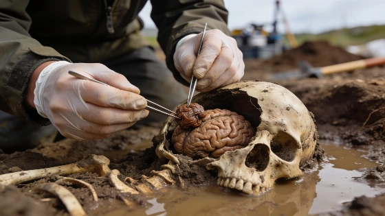
Researchers have identified a chemical process that explains why brain tissue sometimes survives for millennia in archaeological contexts, challenging assumptions about soft tissue decay and offering new insight into ancient preservation
Archaeologists have long noted a paradox in the archaeological record: while most soft tissues rapidly decompose after death, brain tissue has been recovered in a surprisingly high number of ancient human burials, even when the rest of the body has decayed to bone. Recent research published in the Journal of Proteome Research provides a new explanation for this phenomenon, suggesting that brain preservation is not a rare accident but the result of a specific chemical process triggered under certain burial conditions.
Preserved Brains in the Archaeological Record
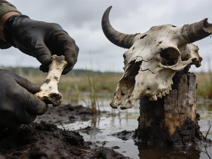
More than 4,400 preserved human brains have been documented from archaeological sites spanning the last 12,000 years. These finds are geographically and chronologically diverse, with many recovered from waterlogged, oxygen-poor environments such as riverbeds, lake margins, and flooded caves. In these contexts, the brain is sometimes the only soft tissue to survive, often appearing as a shrunken, protein-rich mass within otherwise skeletal remains. This selective preservation has challenged conventional understanding of postmortem decay, as waterlogged conditions are typically associated with accelerated decomposition rather than conservation.
Traditional mechanisms for soft tissue preservation-such as mummification, freezing, or saponification-usually affect multiple organs and tissues. However, a significant proportion of archaeological brains do not fit these models, raising questions about the underlying processes. The new study addresses this gap by focusing on the unique chemical and structural properties of brain tissue and the environmental factors that influence its decay.
Experimental Evidence from Animal Models
To investigate the preservation mechanism, researchers conducted controlled experiments using mouse carcasses buried in environments with varying levels of water and oxygen. Over a six-month period, brains were sampled at multiple intervals and analyzed using high-resolution mass spectrometry to track protein degradation and modification. The results revealed that while initial decay processes were similar across conditions, oxygen availability became the critical factor after several weeks. High-oxygen environments led to rapid and extensive protein breakdown, while low-oxygen, waterlogged conditions favored the formation of crosslinked, insoluble protein aggregates that resisted further decay.
This chemical pathway involves free radicals-highly reactive molecules that, in the presence of abundant oxygen, drive a chain reaction leading to complete protein degradation. In hypoxic (low-oxygen) settings, however, the process is interrupted, and intermediate products form stable crosslinks within the brain's protein matrix. The brain's high content of redox-active amino acids, metal ions, and membrane structures makes it particularly susceptible to this self-limiting preservation pathway. The skull may also contribute by restricting oxygen and fluid exchange compared to other tissues.
Implications for Archaeology and Beyond
The identification of this preservation mechanism has significant implications for interpreting ancient human remains. It suggests that the presence of preserved brain tissue in archaeological contexts is not simply a matter of chance, but reflects a reproducible chemical process under specific environmental conditions. This insight may help explain why some burials yield only brain tissue among otherwise skeletonized remains, and why similar preservation is rare in other organs.
In addition to advancing archaeological science, the findings may inform research in other fields. The molecular signature of these decay-resistant brain proteins closely resembles patterns observed in certain neurodegenerative diseases, such as Alzheimer's, raising the possibility that ancient preserved brains could provide new perspectives on disease processes. Further comparative studies of other tissues and burial environments may clarify whether this pathway is unique to brain tissue or more broadly applicable.
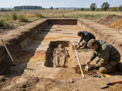
Understanding the chemical and environmental factors that influence preservation is also relevant for interpreting other archaeological discoveries. For example, studies of burial contexts beneath modern structures, such as the investigation of human remains beneath Hermann Göring's former residence at the Wolf's Lair, have highlighted the importance of stratigraphic and taphonomic analysis in reconstructing postmortem histories. For more on how burial environments affect preservation, see this analysis of cemetery remains beneath a Nazi-era structure.
Preservation and Taphonomy Explained
Taphonomy is the study of processes that affect biological remains after death, including decomposition, preservation, and alteration by environmental factors. In archaeological contexts, taphonomic analysis helps researchers distinguish between changes caused by burial conditions and those resulting from human activity or pathology. Factors such as soil chemistry, moisture, temperature, and oxygen availability can dramatically influence which tissues survive and how they are altered. Understanding these processes is essential for interpreting the archaeological record, as preservation bias can affect what evidence is available for study and how past lives are reconstructed.